Track 1: Synapse-Level Connectomics (Flies)
A Tiny but Expansive Brain: The World of the Fly
We often do not think much about flies beyond the hot summer days when they appear around overripe bananas and forgotten fruit. Yet, these tiny insects are far more remarkable than a simple nuisance. Fruit flies possess brains roughly the size of a poppy seed, but within this miniature structure lies an extraordinary capacity for complex behavior.
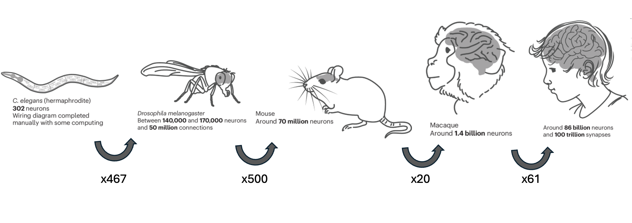
Comparison of brains and neuron numbers. Adapted from Nature Methods. Credit: T. Mitra. Sources: H. Dong, UCLA; M. Helmstaedter, MPI for Brain Research; P. Mitra, CSHL.
Despite their small size, flies can perform a remarkable range of behaviors. They produce courtship songs to attract mates, engage in aggressive encounters with rivals, and execute essential survival behaviors such as locating food, avoiding predators, and responding to changes in light and their environment. The ability of such a small nervous system to generate diverse and sophisticated behaviors makes the fruit fly a powerful model for understanding how neural circuits give rise to behavior.
But how can we understand how a tiny brain produces such complex capabilities? A fundamental step is determining how neurons (the cells responsible for processing and transmitting information) are connected. The pattern of these connections, including which neurons communicate with one another and the strength of those connections, forms the foundation of how information flows through the brain.
This field of mapping neural connections is known as connectomics. Just as a roadmap reveals how cities are connected and how movement occurs between them, a connectome provides a structural blueprint of the brain, showing which neurons are connected and how they communicate.
The field began with a major milestone in the 1980s, when researchers fully mapped the 300-neuron nervous system of a worm, Caenorhabditis elegans, making the first synapse-level connectome detailing how neurons are connected through synapses—the tiny gaps where signals are passed from one neuron to the next.
Now, thanks to advances in microscopy and powerful computational tools, scientists have mapped the neuronal wiring of the fly brain, which contains over 140,000 neurons and more than 50 million synapses. This connectome of Drosophila melanogaster (fruit fly) offers a breathtaking look into how a compact yet complex brain supports vision, movement, memory, and behavior—and now, it’s accessible not just to researchers, but to students and educators around the world.
Over the past few years, researchers at the Janelia Research Campus (part of the Howard Hughes Medical Institute), the MRC Laboratory of Molecular Biology (University of Cambridge), Princeton University, and the Connectomics group at Google have released complete synapse-level connectomes of the entire fruit fly brain and central nervous system (CNS). These datasets capture the full network of neural connections across hundreds of thousands of neurons in both male and female flies representing one of the most detailed neuronal maps ever created.
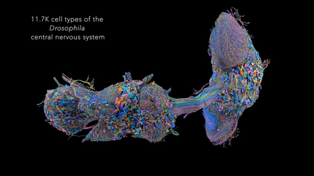
Male CNS Connectome — The complete connectome of the Drosophila male central nervous system (CNS), providing a large-scale map of neuronal connectivity across the brain and ventral nerve cord. Learn more about the Male CNS Connectome · Berg et al., 2025.
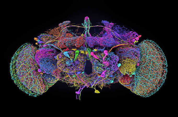
Female FlyWire Connectome — The FlyWire connectome provides a detailed map of neurons and their synaptic connections in the female Drosophila brain. Learn more about FlyWire · Dorkenwald et al., 2024 · Schlegel et al., 2024.
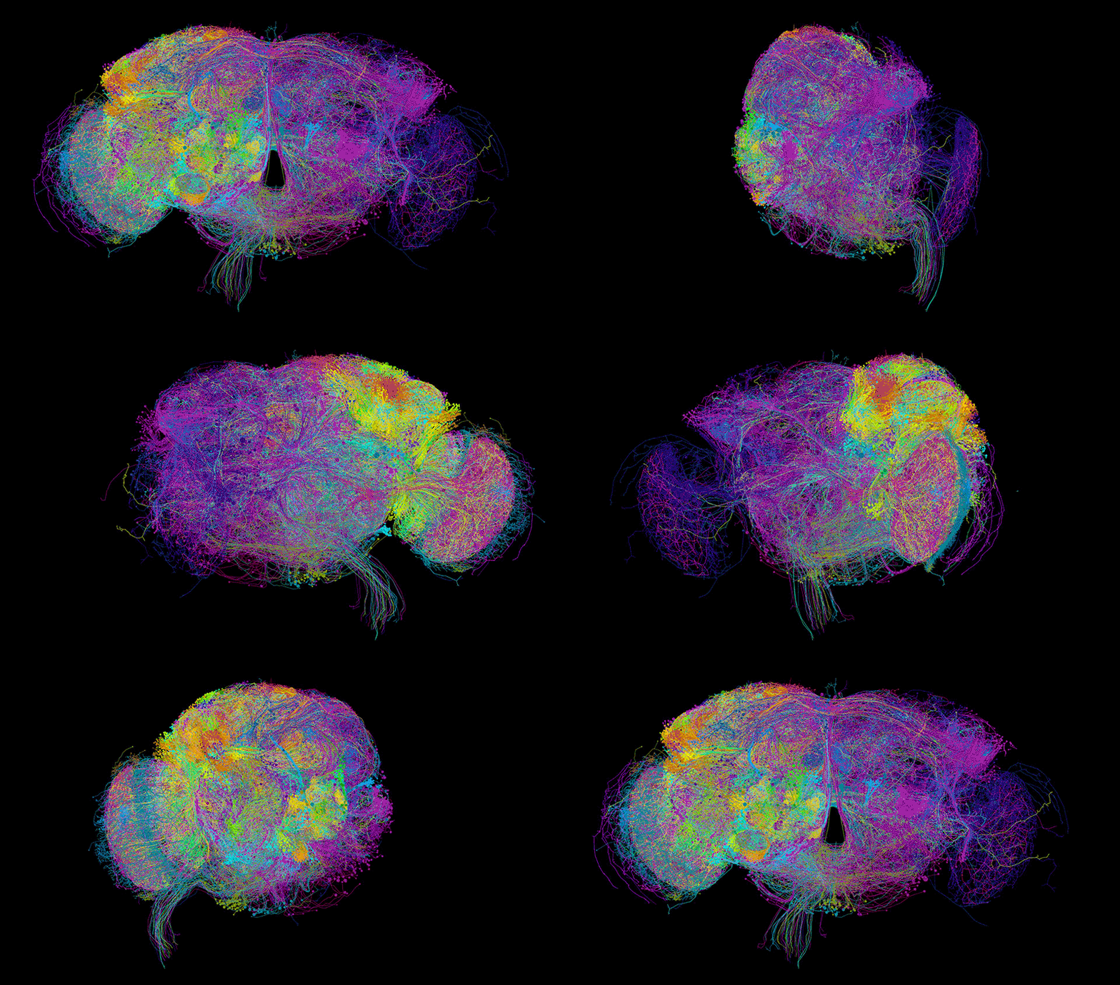
Rendering of FlyWire Neurons — A 3D visualization of neurons reconstructed from the FlyWire connectome.
Larval Connectome — Visualization of the Drosophila larval connectome. Winding et al., 2023.
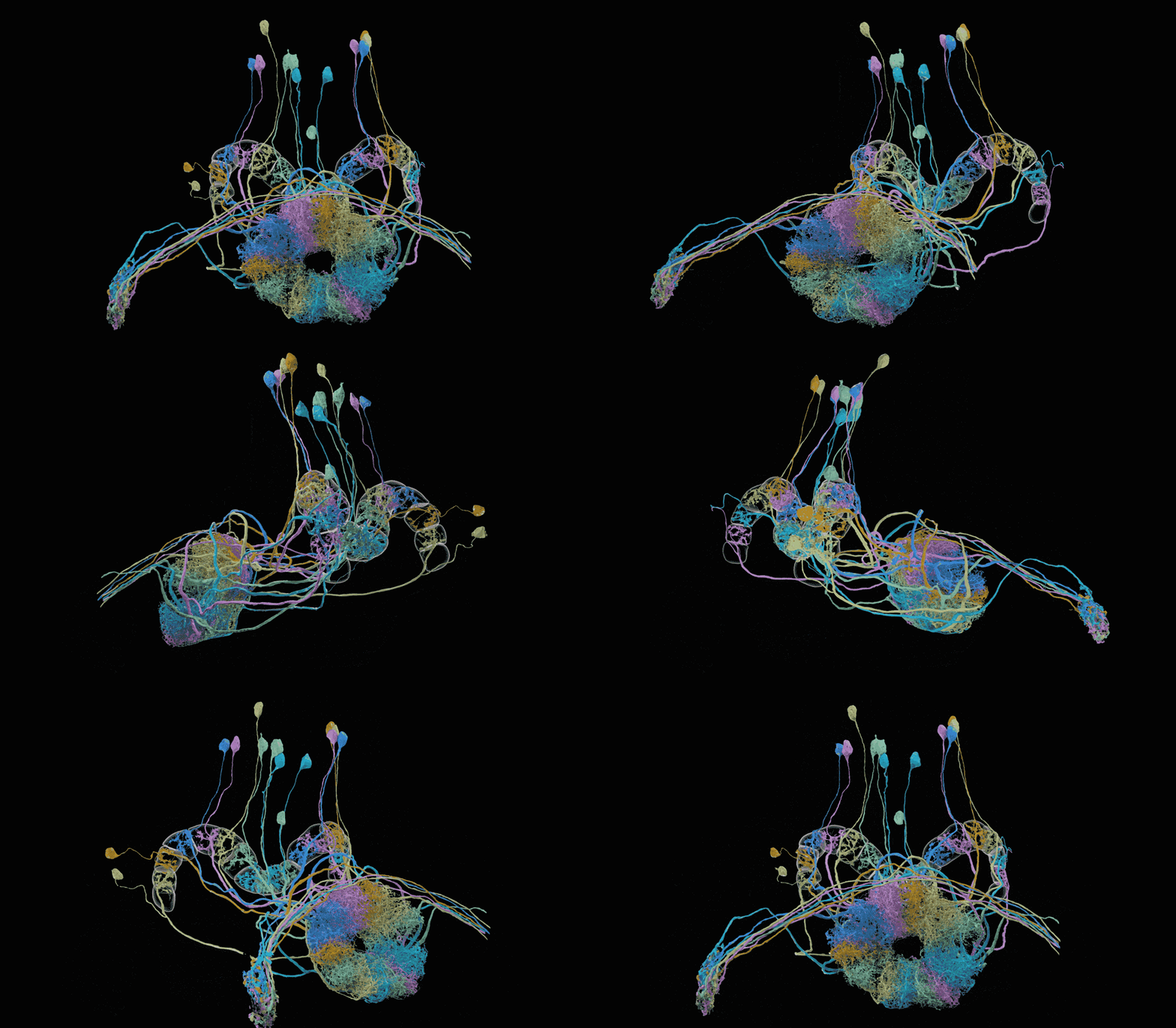
EPG Ring Neurons — 3D visualization of EPG ring neurons in the central complex from the Hemibrain connectome. EPG neurons help the fly keep track of its heading, acting like an internal compass that represents the direction the fly is facing. Learn more about the Hemibrain · Scheffer et al., 2020.
- Dorkenwald, S., Matsliah, A., Sterling, A. R., Schlegel, P., Yu, S., McKellar, C. E., Lin, A., Costa, M., Eichler, K., Yin, Y., Silversmith, W., Schneider-Mizell, C., Jordan, C. S., Brittain, D., Halageri, A., Kuehner, K., Ogedengbe, O., Morey, R., Gager, J., … Zandawala, M. (2024). Neuronal wiring diagram of an adult brain. Nature, 634(8032), 124–138. 10.1038/s41586-024-07558-y
- Schlegel, P., Yin, Y., Bates, A. S., Dorkenwald, S., Eichler, K., Brooks, P., Han, D. S., Gkantia, M., dos Santos, M., Munnelly, E. J., Badalamente, G., Serratosa Capdevila, L., Sane, V. A., Fragniere, A. M. C., Kiassat, L., Pleijzier, M. W., Stürner, T., Tamimi, I. F. M., Dunne, C. R., … Jefferis, G. S. X. E. (2024). Whole-brain annotation and multi-connectome cell typing of Drosophila. Nature, 634(8032), 139–152. 10.1038/s41586-024-07686-5